The CMMT / BCCHR Imaging Core Facility invites you to utilize our: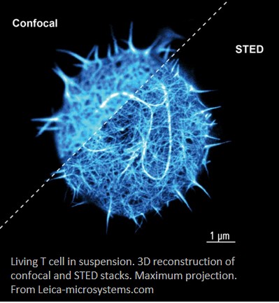
Leica SP8X STED White Light Laser Confocal Imaging system
The system is available to all researchers in our community at the following rates:
- $40/hr for CMMT / BCCHR
- $60/hr for External Academic
- $80/hr for Industry
About Our System
The Leica TCS SP8 STED 3X super-resolution microscope is based on the STED (Stimulated Emission Depletion) principle and is the only purely optical superresolution imaging modality that does not need image processing to generate the image and therefore minimizes the possibility of image artifacts. Image acquisition with STED is much faster than other super resolution methods and the very low energy depletion laser makes it possible to image live cells in the super resolution mode.
The SP8 confocal microscope uses the white light laser for its illumination source. This laser can be set to any wavelength or trillions of laser line combinations. It ensures that there is an appropriate laser line for any fluorophore and most any combination that you may require in your sample.
Our system includes three high sensitivity HyD detectors as well as two GaAsP PMTs. The single step amplification process of the HyD detectors ensures that they are essentially noise free. The combination of high sensitivity and low noise will allow you to image even the most weakly stained samples with minimum exposure to laser light.
The SP8 confocal microscope stand has been configured with a tandem scanner. The FOV scanner allows acquisition of high resolution images with a large field of view. The resonant scanner can be used to image highly dynamic processes with a high temporal resolution.
 The SP8 confocal microscope has a highly efficient light path including a prism-based spectral detection system that is fully tunable and filter-free allowing for true spectral imaging. In grating based spectral detectors a significant amount of light is lost to higher order dispersion and polarization effects. The prism based system in contrast is extremely transparent and nearly all the light emitted by the sample reaches the detector.
The Leica SP8 confocal microscope also features the AOBS beam splitter. As opposed to traditional fixed glass based dichroic beam splitters, the AOBS is a fully tunable beam splitter that makes the microscope more flexible and sensitive. Because of this device you can simultaneously image dye combinations such as GFP-YFP or CFP-GFP that are not possible with other microscopes.
The superresolution confocal microscope is based on the new DMi8 inverted microscope. It is fully automated and has the added hardware based closed loop focus drift compensation system. In addition to the focus drive, it includes an additional high resolution fast focus drive that allows the microscope to correct for drift faster and more precisely. In addition the camera port on the new DMi8 microscope has a larger field of view suited for the large chip of the sCMOS cameras.
System Specifications

(all links are to additional information on vendor sites or youtube)
Leica SP8X STED White Light Laser Confocal Microscope
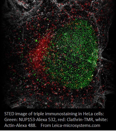
- Super-resolution gated STED imaging down to sub-50nm sub-cellular structures
- Complete spectral freedom
- Up to 8 freely tunable laser lines simultaneously from 470 to 670nm White Light Laser, 1.5 mw, in steps of 1nm, pulsed at 80 MHz
- 50 mw 405 diode laser
- 592 nm STED depletion laser line output > 1.5 W
- AOBS (Acousto-Optical Beam Splitter)
- Two scanners mounted on Tandem scanner
- Conventional Scanner scans 1-1800 lines/s for high spatial resolution with largest field of view
- Resonant Scanner scans at fixed 8000 lines/s for high temporal resolution
- Objectives:
- 10X / 0.4 HC PL APO CS, WD 2.2 mm
- 20X / 0.75 HC PL APO CS2, WD 0.62 mm
- 40X / 0.85 HC PL APO CS, WD 0.21 mm, correction collar for 0.11-0.23 mm coverslip, not for 405 nm excitation
- 63X / 1.3 Glycerol HC PL APO CS2, WD 0.3 mm, correction collar for 0.14-0.19 mm coverslip, 20-40° C
- 100X / 1.4 Oil HC PL APO CS2 STED, WD 0.13 mm
- Stage
- SuperZ Galvo, Galvo Flow sweeping through live specimens in 4D
- Adaptive Focus Control(AFC) actively and dynamically regulates the focus position
- Stage inserts for slide, petri dish up to 35 mm, labtek chamber, and multi-well plate
- Stage Top Incubator for heating, moisture and premixed gas (5% CO2/95% Air) flow control for live cell imaging
- 5-Channel Leica SP Spectral Fluorescence Detection, three HyD and two regular PMT detectors, 400-800nm, minimum 5nm
- HyD detector
- Supersensitive photon detection with maximum quantum efficiency of ~45 % at 530 nm (twice as much as a standard PMT)
- Very low dark noise to render the finest details
- Photon counting capability for fluorescence detection
- One Transmitted light detector for bright field, or DIC imaging
- FRET AB, FRET SE, FRAP, FRAP XT
- FRAP Wizard
- Efficient bleaching by optional Zoom in and change of format during bleach
- Fly Mode for fast recording of recovery by bleaching and dada readout in one frame
- xyzt mode for pre- and post-bleach acquisition for 3D FRAP analysis
- Online and offline quantification of data
- FRET Acceptor-Photobleaching Wizard
- FRET measurements with fixed and immobile samples
- FRET efficiency calculated for user-defined regions
- Results displaced as FRET efficiency map
- FRET Sensitized Emission Wizard
- Correction of background fluorescence and cross-talk
- Online and offline quantification of FRET efficiency in user-defined regions
- Result displayed as FRET efficiency map
- FRAP Wizard
- Full spectral characterization of images, l-l scan of excitation-emission spectral image map
- LightGate
- Zero background by removing reflection and autofluorescence by adjustable detection time gate
- Removing non-wanted fluorescence to increase image contrast
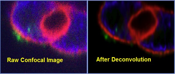
- Huygens Professional Deconvolution Software Package restores STED/Confocal images back to original objects through mathematical de-blurring and de-noising. It enhances image resolution and signal/noise ratio, and removes noise background.
- Wide-field Fluorescence Imaging
- ORCA-Flash4.0 V2 Digital CMOS Camera (High-end) for fast live imaging
- 80% QE at 600nm
- 2048 X 2048 resolution, 6.5 μm2 pixel, 30 frames/s, 16 bit data output
- full well 30, 000 e–, read noise 1.6 e– rms (1.0 e– median), dark current 0.06 e–/pixel/s
- Filter cubes
- DAPI: EX: BP 350/50, EM: BP 460/50
- GFP: EX: BP 470/40, DM 500, EM: BP 525/50
- RFP: EX: BP 560/40, DM 595, EM: BP 630/75
- Triple D/F/TX-S: EX BP 420/30, 495/15, 570/20; DM 415, 510, 590; EM: BP 465/20, 530/30, 640/40
- MetaMorph Premier Imaging Acquisition Software
- Multi-channel, multi-site, and high-throughput time-lapse fluorescence and BF/DIC imaging
- ORCA-Flash4.0 V2 Digital CMOS Camera (High-end) for fast live imaging
Billing Authorization and Invoicing
Invoicing will be processed on a monthly basis by the Workday Internal Service Delivery (ISD) process for UBC customers. To enable monthly billing to your UBC grant, please complete the ISD Authorization Form available here. Non-UBC customers will be invoiced monthly.
Contact Us
For additional information and for system training:
Mr. Jingsong Wang
(604) 875-2000 ext.7564
imaging@cmmt.ubc.ca
For billing information and for further information on CMMT Core Facilities:
Michael Hockertz
hockertz@cmmt.ubc.ca
(604) 875-3816